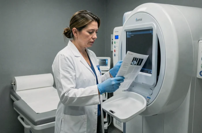
Bone densitometry remains the cornerstone of osteoporosis diagnosis, but the way this examination is performed and interpreted is evolving rapidly. In Aix-en-Provence, several medical imaging centers offer this measurement of bone mineral density, primarily through dual-energy X-ray absorptiometry (DXA). Recent innovations challenge the exclusive role of the T-score and pave the way for complementary approaches, both technologically and clinically.
Beyond the T-score: Bone Quality as a New Screening Criterion
For years, the T-score has served as the almost sole reference for assessing fracture risk. This figure, derived from the comparison between the patient’s bone density and that of a healthy young adult, has a known limitation: a normal density does not exclude a high fracture risk.
Tools like TBS Reveal (developed by Medimaps) and BoneScore aim to fill this gap. Their approach is based on the analysis of bone quality, meaning the microarchitecture of bone tissue, rather than just its raw density. TBS Reveal utilizes already acquired DXA images to extract a trabecular texture score, without additional radiation or extra examination.
The shift towards a comprehensive assessment of bone strength changes how physicians interpret results. A patient whose T-score remains in the osteopenia range may present a significant fracture risk if the quality of their bone tissue is compromised. Conversely, decreased density with good microarchitecture does not necessarily warrant the same treatment.
In practice, bone densitometry in Aix-en-Provence is part of a health hub where medical imaging is diversifying. Centers that adopt these complementary tools offer a more nuanced reading of risk, even though their dissemination remains gradual in local facilities.
X-ray Free Bone Densitometry: Where is REMS Technology Now?
DXA uses ionizing X-rays. The dose received by the patient is low, but the issue of radiation arises when regular follow-up is necessary, especially in patients undergoing long-term treatment. The CDC reminds that each DXA examination exposes to radiation, and the benefits must be weighed against the risks in a context of repetition.
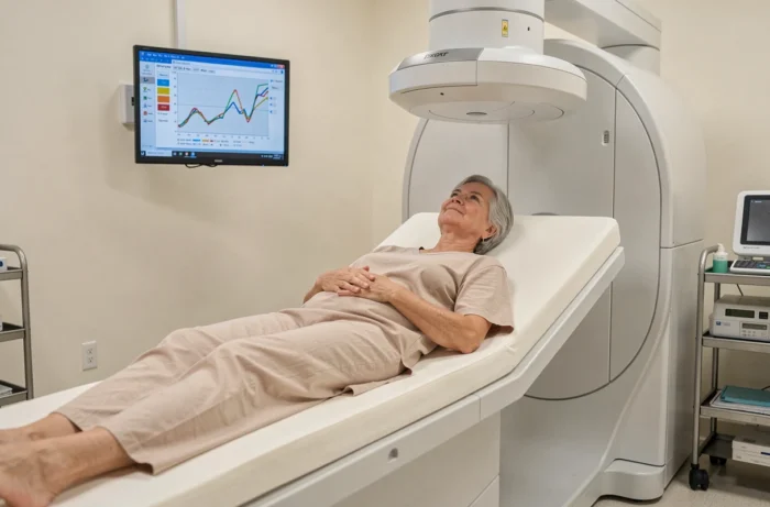
The REMS (Radiofrequency Echographic Multi Spectrometry) technology, developed by Echolight, offers an alternative. This portable device operates using ultrasound and radiofrequency, without any radiation. It assesses bone density, fragility, and even body composition.
For patients requiring frequent monitoring (long-term corticosteroid therapy, hormone replacement therapy, chronic diseases affecting bone metabolism), a radiation-free examination allows for closer follow-ups. The available data do not yet allow for conclusions about the exact equivalence between REMS and DXA measurements in all clinical contexts. Field feedback varies on this point depending on the studied populations.
What REMS Technology Changes Practically
- The portability of the device allows the examination to be performed outside of radiology departments, including in rheumatology or general medicine consultations
- The absence of radiation permits follow-ups at shorter intervals without concerns about cumulative dose
- The simultaneous assessment of body composition (fat mass, lean mass) adds a metabolic dimension to the bone assessment
This technology is not yet available in all centers in Aix-en-Provence, but it illustrates a broader movement towards less burdensome and more accessible examinations.
Osteoporosis Screening Pathway: At-Risk Profiles at the Limits of the System
French screening recommendations target specific profiles: postmenopausal women with risk factors (history of fracture, corticosteroid therapy, low body mass index), men over seventy, patients on treatments that induce bone loss. Systematic screening of the general population is not recommended.
This targeting logic has its virtues (avoiding unnecessary examinations) and its blind spots. Patients without identified risk factors but with silent osteoporosis go under the radar until the first fracture. The CDC also emphasizes that DXA is used to monitor changes in bone density, whether it improves, remains stable, or worsens, and not just to make an initial diagnosis.
Clinical Situations Justifying Regular Follow-Up
- Hormone replacement therapy in postmenopausal women: the examination checks the effectiveness of the treatment on bone mass at the lumbar spine and femur
- Long-term corticosteroid therapy: bone loss can be rapid and requires close monitoring
- Endocrine disorders (hyperparathyroidism, hyperthyroidism): the bone impact must be quantified and monitored over time
- History of fragility fracture: initial screening is supplemented by follow-up to adapt the therapeutic strategy
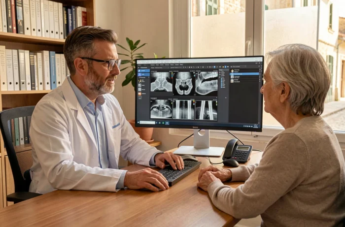
In Aix-en-Provence, medical imaging centers recommend conducting follow-up examinations in the same facility and on the same machine to ensure the comparability of measurements from year to year. A change of machine can introduce variations that distort the interpretation of progress.
Bone Densitometry and Fracture Risk Prediction: The Limits to Know
The question of the predictive reliability of bone densitometry is a subject of discussion in the medical literature. Bone density alone predicts only part of the fracture risk. Other factors come into play: age, personal and family history, fall risk, ongoing treatments.
Complementary tools (TBS, FRAX) attempt to integrate these parameters into an overall score. However, their systematic use in everyday practice remains uneven across centers and practitioners. Some radiologists incorporate them into reports, while others stick to the classic T-score and Z-score.
The development of technologies like REMS or bone texture analysis does not eliminate this complexity. It shifts it by multiplying the data available to the clinician, which requires appropriate training and updated interpretation protocols.
The challenge for the coming years, in Aix-en-Provence and elsewhere, lies less in the availability of devices than in the ability of care pathways to integrate these new data into individualized medical decision-making. Technology is advancing faster than field practices, and it is in this gap that the real quality of osteoporosis screening and follow-up is at stake.